Physician Led • Outcomes Centered • Patient Focused

Tenex Procedure for Foot or Ankle

Is foot or ankle pain keeping you from doing what you want? Don’t let your pain keep you on the inside looking out.


At Stadia Sports Medicine, we are performing a revolutionary procedure that is changing the lives of our patients for the better.
Tenex Health TX offers a safe, quick, minimally invasive solution to relieve pain, restore mobility, and rapidly return patients to their normal activities. The procedure is performed in less than 20 minutes, patients are awake and alert throughout (only a local anesthetic is used), and no sutures or stitches are needed—only a small adhesive bandage is required. Most patients are able to return to normal activity within 6-8 weeks. Call us at 515.221.1102 or email us at reception@stadiasportsmedicine.com today for more information about Tenex Health TX. Maybe it’s time to move forward and leave the pain behind.
Dr. Carlson was one of the first doctors in our area to be trained on this groundbreaking technology.

Is your pain interfering with your work, your play or simply living your life?

Are you silently suffering because you were previously told there is no other option for you but a major or invasive surgery with a long recovery?  Or perhaps you know that all the ice and stretching is not going to make your pain disappear?
If you have tried physical therapy, cortisone injections, medication, or just taking time to ice, stretch and rest and are still in pain, talk to us!  We now have a solution that does not involve general or open surgery, may give you quick pain reduction and should have you back to enjoying the things you love in a few weeks to a few months.
We CAN help!
Stadia Sports Medicine practice offers Tenex Health TX™, a safe, quick procedure specially designed for those who are suffering from painful conditions associated with tendon damage, commonly referred to as pain in the rotator cuff, Tennis or Golfer’s elbow, Runner’s or Jumper’s knee, Achilles tendonitis or Plantar fasciitis.
The Tenex Health TX System is based on advanced technology developed in collaboration with the world-renowned Mayo Clinic.  It combines ultrasound imaging (which is also used to see a baby in the womb) to identify damaged tissue with the advanced TX MicroTip™ which precisely breaks down and removes only the damaged tissue that causes pain.  Removing the damaged tissue allows the tendon to heal and provides pain relief.  The Tenex Health TX procedure is not open surgery and can be performed in an office or procedure room in an outpatient surgery center.
Tenex Health TX benefits:
  • Quick pain relief
  • Rapid return to normal activities
  • Local anesthetic used instead of general anesthesia
  • No sutures, no stitches (requires only a small, adhesive bandage)
  • Minimally invasive, not open surgery
  • Covered by most insurances

We are here for you…

You don’t have to live with pain!  We are happy to discuss Tenex Health TX or any other option that may work for you.  Please call our office at 515-221-1102 to schedule a consultation.   You can also find more information on the procedure at https://tenexhealth.com/.

Questions and Answers:

Q:  What is a tendon?
A: A tendon is a strong, yet flexible band of tissue that connects muscle to the bones around your joints.  Tendons and muscles work together to create a pulling force that enables you to move your joints.  This allows you to bend, walk, jump and move in many different ways.  When tendons are damaged, movement in a joint can be very painful and limit your range of motion.
Q:  What causes tendon pain?
A: Tendons are injured by either outright trauma or by repetitive injury (damage to the tendon from overuse in work, exercise or activity).  When the tendon is traumatized, it tries to heal itself, but often eventually forms a scar.  This scarred tendon causes pain.  Eventually, scar tissue in the tendon will limit your ability to move the joint and do the activities that you need or like to do.  If you don’t treat the damaged tendon, the pain in your joint will likely become worse over time.
Q:  What are the most common types of tendon injury?
A: The most commonly injured tendons include:
  • Achilles tendonitis
  • Patellar tendonitis (“Jumper’s knee”)
  • Plantar fasciitis
  • Rotator cuff tendonitis
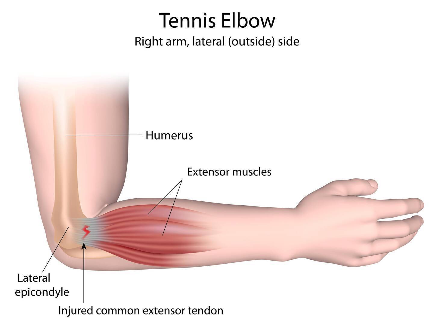
  • Tennis elbow
  • Golfer’s elbow
Q:  What are the options for treatment of chronic tendon problems?
A: At Stadia Sports Medicine, the following options are offered:
  • Cutting-edge Physical Therapy
    • Stadia Sports Medicine was the first clinic in Des Moines to utilize the ASTYM technique with our patients, having used it since our opening in 2006.  ASTYM was developed by Dr. Carlson’s fellowship director at Ball State University in Muncie, Indiana, Tom Sevier, M.D., who went on to found Performance Dynamics
      –the company that trains clinicians in the ASTYM technique.  Thus, practitioners at Stadia have experience
      with ASTYM going back to the 1990’s, and our therapy staff has a broader depth of experience with this technique than any other physical therapy group in Des Moines.
    • Stadia Sports Medicine’s physical therapists also utilize the latest in therapy techniques designed to speed tendon healing and promote more rapid recovery from injury. We do not rely on techniques such as electrical stimulation or therapeutic ultrasound which are commonly used in therapy settings but which
      have not been shown to be effective for these problems.  Our approaches to patients are grounded in
      the medical literature.
  • Injection therapies – Injection treatment can take one of several different approaches, including:
    • Steroid injections – Steroid injections reduce pain and swelling associated with chronic tendon injury.  Although steroid cannot be directly injected into tendons, it can be injected around the tendons themselves in order to provide pain relief.  This is best done under guidance, both to ensure a good response and to minimize procedural pain.  Steroid injections can assist the rehabilitation process.  They are covered by almost all insurance plans.
    • Prolotherapy – Prolotherapy involves the injection of an irritant substance into an injured tendon with the goal of stimulating a healing response.  Although different substances can be used, at Stadia, we utilize Dextrose – a form of sugar – because of its safety profile and use in studies that have shown a clinical
      benefit to patients.  In the case of prolotherapy, dextrose is injected directly into the tendon in the area of
      injury, with guidance utilized to promote precision of placement.  Although prolotherapy is not a covered
      benefit for most insurance plans, it’s low expense makes it a feasible option for most patients.
    • PRP (Platelet-rich plasma) injections – PRP involves the injection of a patient’s own blood into an area of tendon (or joint) injury.  Specifically, the blood is prepared prior to injection by being spun in a centrifuge so that the platelets can be isolated for injection. Platelets contain many factors in them that promote wound and tissue healing.  PRP shows promise as a useful therapy when more conservative measures have failed.  At Stadia we utilize the Harvest PRP system, which provides an optimal concentration of platelets for injection to the injured area – something that has been shown to be important for optimal response.  We also utilize guidance for all PRP injections, to increase the likelihood of a good response to treatment.  PRP is not a covered benefit for most insurance plans and is usually considered a cash service, payable at the time of the procedure.
    • Percutaneous tenotomy – Click to find out more about this treatment.
    • Surgical tenotomy – Used to remove the scar tissue, surgery has been found to be beneficial.  However, it is accompanied with potential side effects of general surgery, unintended damage to surrounding muscle and
      tissue and a lengthy recovery period with restricted activity.
Q:  What is percutaneous tenotomy?
A: Percutaneous tenotomy is a procedure that can be used to help heal chronic tendon pain that is not getting better with
traditional non-operative measures.  It involves the removal of scarred tissue from an injured tendon and can be thought of as a bridge between traditional surgical tenotomy and these non-operative treatments. This short video outlines the procedure:
Q:  Does percutaneous tenotomy work?
A: Yes.
Outcomes seem to mimic that of surgical treatment, with less post-procedure pain and more rapid recovery, as well as lower overall costs.  There is a reported 80-90% success rate among patients not responding to conservative therapies.
Q:  What happens during a percutaneous tenotomy procedure?
A: Percutaneous tenotomy is done entirely under local anesthesia.  Ultrasound guidance technology (the same equipment used to look at a baby in the womb) is first used to identify the area of tendon injury, and then appropriate skin markings are made.  Following this, using a small needle and ultrasound guidance, local anesthetic is injected into and around the area of tendon injury.  Once the area is anesthetized, a special tool is inserted into the scarred tissue.  This tool delivers ultrasonic energy that is specifically designed to break up and remove damaged tissue safely and quickly, without disturbing the surrounding healthy tendon tissue.  Total treatment time for this procedure averages 15-20 minutes.  After the scar tissue is removed, the tiny opening is closed with a steri-strip.
Q:  What is the recovery like from percutaneous tenotomy?
A: Following the procedure, patients are discharged to home with over-the-counter pain relievers usually adequate to
control post-procedure pain.  Restrictions following this procedure are going to be individualized, but general guidelines are as follows:
  • Tennis/golfer’s elbow
    • Light activity for the first week
    • Begin gentle strengthening at two weeks
    • Begin more aggressive strengthening and lifting around four weeks post-procedure
  • Patellar tendonitis/Quadriceps tendonitis
    • Crutch protection for the first week
    • Normal weight-bearing between weeks 2-4
    • Progress into monitored exercise starting at four weeks post-procedure
  • Achilles tendonitis
    • Walking boot for first two weeks
    • Normal daily walking between weeks 2-4
    • Progress into monitored exercise starting at four weeks post-procedure
  • Plantar fasciitis
    • Walking boot for first 1-2 weeks
    • Normal daily walking between weeks 2-4
    • Progress into monitored exercise starting at four weeks post-procedure.
  • Calcific rotator cuff tendonitis
    • Sling for comfort for the first week
    • Modified lifting/use for weeks 2-4
    • Progress into monitored exercise starting at four weeks post-procedure
Q:  Is percutaneous tenotomy safe?
A: Yes.
With thousands of procedures performed in the United States, there have been no reports of significant complications to date.
Q:  Is percutaneous tenotomy covered by insurance?
A: Yes.
Percutaneous tenotomy is a covered benefit on most insurance plans.
Q:  What studies have looked at this treatment?
A: Summarized below are several studies looking at the effectiveness of percutaneous tenotomy for certain medical
conditions:
  • Author: Koh, Joyce.
  • Condition: Tennis Elbow
    • 20 patients with chronic tennis elbow who had failed non-surgical treatment
    • Post-treatment guidelines:  Two weeks of activity modifications
    • Follow-up at 2, 4, 12, 24 and 52 weeks post-procedure
      • 19/20 patients were pain-free
      • No complications
      • Normalized appearance on ultrasound
  • Author: Patel
    • Condition: Plantar fasciitis
    • 12 patients with symptoms of heel pain for >
      four months

      • 11/12 were pain-free by six weeks and remained
        pain-free at 3 and 6 months
  • Author: Elattrache
    • Condition: Patellar tendonitis
    • 16 patients with symptoms for at least four
      months, all having failed conservative treatment
    • 10 collegiate-level athletes
      • 15/16 were pain-free by six weeks and remained
        pain-free at 3 and 6 months.
      • All ten athletes returned to their prior level
        of competition within three months.
Q:  What is the cash price for percutaneous tenotomy?
A: The cash price varies slightly by location, but averages around $1700, which includes post-procedure
visits during the first 90 days.

Posted in Athletes / Patients, General, Services | Tagged , , , , , , , , , , , , | Leave a comment

I have elbow pain, what can I do about it?

Is elbow pain keeping you from simple everyday tasks like drinking coffee, lifting a bag or driving? Don’t let your pain keep you down.

At Stadia Sports Medicine, we are performing a revolutionary procedure that is changing the lives of our patients for the better.
Tenex Health TX offers a safe, quick, minimally invasive solution to relieve pain, restore mobility, and rapidly return
patients to their normal activities. The procedure is performed in less than 20 minutes, patients are awake and alert
throughout (only a local anesthetic is used), and no sutures or stitches are needed—only a small adhesive bandage is
required. Most patients are able to return to normal activity within 2-6 weeks. Call or write to us today for more
information about Tenex Health TX. Maybe it’s time to move forward and leave the pain behind.
Dr. Carlson was one of the first doctors in our area to be trained on this groundbreaking technology.

Tennis elbow is one of the most common types of tendon injury. Is your elbow pain interfering with your work, your play or simply living your life?

Are you silently suffering because you were previously told there is no other option for you but a major or invasive surgery with a long recovery?  Or perhaps you know that all the ice and stretching is not going to make your pain disappear?

If you have tried physical therapy, cortisone injections, medication, or just taking time to ice, stretch and rest and are still in pain, talk to us!  We have a solution that does not involve general or open surgery, may give you quick pain reduction and should have you back to enjoying the things you love in a few weeks to a few months.

We CAN help!

Stadia Sports Medicine practice offers Tenex Health TX™, a safe, quick procedure specially designed for those who are suffering from painful conditions associated with tendon damage, commonly referred to as pain in the rotator cuff, Tennis or Golfer’s elbow, Runner’s or Jumper’s knee, Achilles tendonitis or Plantar fasciitis.

The Tenex Health TX System is based on advanced technology developed in collaboration with the world-renowned Mayo Clinic.  It combines ultrasound imaging (which is also used to see a baby in the womb) to identify damaged tissue with the advanced TX MicroTip™ which precisely breaks down and removes only the damaged tissue that causes pain.  Removing the damaged tissue allows the tendon to heal and provides pain relief.  The Tenex Health TX procedure is not open surgery and can be performed in an office or procedure room in an outpatient surgery center.

Tenex Health TX benefits:

  • Quick pain relief
  • Rapid return to normal activities
  • Local anesthetic used instead of general anesthesia
  • No sutures, no stitches (requires only a small, adhesive bandage)
  • Minimally invasive, not open surgery
  • Covered by most insurances

We are here for you…

You don’t have to live with pain!  We are happy to discuss Tenex Health TX or any other option that may work for you.  Please call our office at 515-221-1102 to schedule a consultation.   You can also find more information on the procedure at http://www.tenexhealth.com/.

Q:  What is the recovery like from percutaneous tenotomy?

A: Following the procedure, patients are discharged to home with over-the-counter pain relievers usually adequate to control post-procedure pain.  Restrictions following this procedure are going to be individualized, but general guidelines are as follows:

  • Tennis/golfer’s elbow
    • Light activity for the first week
    • Begin gentle strengthening at two weeks
    • Begin more aggressive strengthening and lifting around four weeks post-procedure

Q:  What studies have looked at this treatment?

A: Summarized below are several studies looking at the effectiveness of percutaneous tenotomy for certain medical conditions:

  • Author: Koh, Joyce.
  • Condition: Tennis Elbow
    • 20 patients with chronic tennis elbow who had failed non-surgical treatment
    • Post-treatment guidelines:  Two weeks of activity modifications
    • Follow-up at 2, 4, 12, 24 and 52 weeks post-procedure
      • 19/20 patients were pain-free
      • No complications
      • Normalized appearance on ultrasound

Here’s a quick animation of how the procedure works:


Click here to find out more!

#Tenex #tenniselbow #percutaneoustenotomy #golferselbow #stadiatenex #tendon #tendonitis #ultrasound #tennis #golf #elbow

Tenex Health TX Video Animation of Procedure


Posted in Athletes / Patients, General, Services | Tagged , , , , , , , , , , , | Leave a comment

Journal Club Review: The use of orthotics to control leg and knee pain with activity

 Mills K, Blanch P, Dev P, Martin M, Vicenzino.  A Randomized Control Trial of Short-Term Efficacy of In-Shoe Foot Orthoses Compared With a Wait-and-See Policy for Anterior Knee Pain and the Role of Foot Mobility. Br J Sports Med 2012;46:247-252.
Hirschmuller A, Baur H, Muller S, Helwig P, Dickhuth H-H, Mayer F.  Clinical Effectiveness of Customized Sport Shoe Orthoses for Overuse Injuries in Runners:  A Randomized Controlled Study.  Br J Sports Medicine 2011;45:959-965.

Custom arch supports can be costly, and people spend a lot of money (millions) on them each year.  Do they work though?  Previous recent studies1,2 suggest that they do, and two studies published in the British Journal of Sports Medicine over the last year reinforce this.

In a study out of Australia published this year, investigators evaluated the effect of orthotics in patients with anterior knee pain (pain coming from the kneecap, or patella).  In this study, forty patients with anterior knee pain were randomized to run in shoes with orthotics or shoes alone (the control group).  No additional treatment was prescribed so that a true treatment effect could be identified.  Patients selected for this study had to be between the ages of 18 and 40, have had pain with activity that was not caused by injury for longer than six weeks and an examination by a physician that met appropriate criteria.  Patients also had to meet two of the following criteria, which have been previously identified as being associated with a higher likelihood of treatment success:

  • A measurable tendency for increased foot width when moving from non-weight-bearing to standing
  • Pain severity of low to moderate degree
  • Age older than 25 years
  • Height shorter than 165 cm (5 feet, 5 inches).

Patients with hip, pelvis or back pain, prior knee damage or patellar tendonitis, prior lower extremity surgery, a history of physical therapy for knee pain in the last three years or with foot conditions that would prohibit use of orthotics were excluded.

Each patient was asked to fill out various validated pain and function surveys at baseline.  Subjects were tested on a treadmill with orthotics of varying firmness to find the most comfortable insert for them.  Both the orthotic and control groups were brought back at the end of six weeks and re-evaluated.

Patients with anterior knee pain who had been issued orthotics had statistically greater improvement in function than the control group, with 79% noting “improvement” or “marked improvement” and nearly half of the group (47.4%) noting “marked improvement.”  Patients with foot widening upon standing experienced a “marked improvement” in 78% of cases.  Only 1/19 control subjects perceived relief by the end of the study period.

This study is helpful because it would suggest that patients with anterior knee pain have a 50% chance of obtaining good pain relief from nothing more than the use of appropriate orthotics.  In other words, one patient for every two patients being treated should get good relief.  If the patient is clinically examined and an effort is made to determine if the patient has a foot type that would potentially respond better, the odds of effective treatment from orthotics would seem to improve even more.  One drawback to this study is that it was not blinded to the patient, meaning that all patients knew whether they were being treated or not, which introduces the potential for placebo effect.


Our second study, this one from Germany, attempted to look at the effectiveness of orthotics for runners with lower limb overuse injuries (shin splints, Achilles tendonitis, Plantar fasciitis, anterior knee pain, IT band syndrome, etc).  The most common diagnosis in this group was Achilles tendonitis, followed by patellar tendonitis.  Runners between 18 and 60 years of age with symptoms in one leg and a history of running over twenty miles per week were offered to participate in the study, assuming no history of lower extremity or spine surgery, recent traumatic injury or ongoing therapies for the current complaint within the last six months.

Patients in this study also were asked to fill out several surveys validated to measure pain and function.  Each participant was given a training diary to complete and was asked to document distance, duration and training intensity for each run session.  Functional surveys were filled out at the end of each week.  Data was collected for two weeks, at which time, groups were randomized to run in either generic orthotics or their standard running shoe.  Orthotic compliance was monitored, with use in at least 80% of all training sessions required.  After eight weeks of treatment, patients were re-evaluated.

Analysis of data showed that there was a statistical improvement in the orthotic group over the course of the study, with measurable improvements in both disability and pain.  Patients in the control group had significant increases in pain and disability over the treatment period, with no ill effects relating to the use of the orthotics.  Of note, there was a slight temporary increase in symptoms in the orthotic group during the first week of the study that reversed by week two and then continued to trend toward improvement.  This emphasizes the importance of laying out proper expectations for patients to minimize the likelihood that they will stop using their orthotics too soon.

Thus, it would appear that orthotics do offer a significant likelihood of pain relief for commonly painful problems involving the legs and feet.  A quick check of publically-available data shows that it is not uncommon for Good Feet Store franchises to have annual revenues of between $500,000 and one million dollars, with patients spending upwards of $1000 for orthotics.  That’s a lot of money that people are shelling out in an effort to control foot or leg pain. We think that’s crazy.  At Stadia, our custom orthotics are priced at $200* and always comes with a no-question return policy of 45 days from the time of the order or about 35 days from the time that the orthotics arrive.  No appointment is necessary to order these either, though if you are struggling with pain, we would always suggest that a physician evaluation is wise.


1Barton CJ. The immediate effects of foot orthoses on functional performance in individuals with patellofemoral pain syndrome. Br J Sports Med 2011;45:193-7.

2Collins N. Foot orthoses and physiotherapy in the treatment of patellofemoral pain syndrome:  randomized clinical trial. BMJ 2008;337:a1735.

*Price subject to change due to a change in our costs.

Click either of these for more info on orthotics offered at Stadia Sports Medicine:

Posted in Sports Medicine Research | Tagged , , , , , , , | Comments Off on Journal Club Review: The use of orthotics to control leg and knee pain with activity

Journal Club Review: Exercise Therapy for Anterior Knee Pain Provides Cost Savings

 
                Exercise Therapy for Patellofemoral Pain Syndrome Costs Society Less Than Usual Care. Tan SS et al. J Med Sci Sports. 2010;20:568-579.

This study, done in the Netherlands on patients with anterior knee pain, showed an overall cost-effectiveness of exercise therapy when compared with usual care of medication and observation.  131 subjects between the ages of 14 and 40 with symptoms between two months and two years were included.  None of the subjects had undergone prior exercise therapy, prior knee surgery or had other known knee problems.  Subjects were randomized to either a six week exercise protocol involving information on anterior knee pain and nine therapy visits or a control group given the information alone.  Total treatment costs, including therapy, physician visits, bracing, imaging studies, medication and work-related costs were monitored in the two groups over a one year observation period.

Although up-front costs in the therapy group were higher than the observation group, when productivity costs were factored in, total costs in the therapy group was less.  In addition, patients in the treatment group obtained better control of their symptoms.

I’ve included this study for one reason.  Personally, I am not that impressed with the cost savings reported in this study.  The total costs for the exercise group were estimated at $1348 versus $1555 for the group that was managed with usual care.  The observation period in this study was only one year, and we don’t have any idea what the long-term trends of both groups look like.  The up-front costs of the therapy group were greater than the usual care group, though outcomes were better in the former group.  I include this study because I think that it highlights our usual approach to anterior knee pain quite well.  At Stadia, when a patient is evaluated by a physician for anterior knee pain, the approach is a focus on effective but cost-conscious care.  Unless the patient is requesting them, x-rays are usually not obtained at the first visit unless arthritis is considered a risk or there are other reasons, (such as swelling in the knees) to obtain them.  Medications are rarely used.  Instead, each patient is offered a home exercise program tailored to their own unique exam.  This program is put onto paper and reviewed with the patient prior to discharge, and the patient is scheduled for a follow-up visit – usually at three weeks from the first appointment.  If, as expected, the patient is improving, their home exercise program is advanced and they are usually discharged with instructions to follow-up if symptoms are not completely resolved within 3-4 weeks.  If struggling at follow-up, x-rays are obtained and other treatment options can be discussed.  Braces are rarely required.  Although we do not deny patients the option of medication or supervised therapy for anterior knee pain (recognizing that patients come to us with different priorities, abilities to follow-through with home exercise or timelines that may require more rapid improvement), we have found that this approach results in significant improvement in the majority of our patients, without the need for physical therapy and without the necessity of imposing prolonged restriction from activity on patients who are seeing us in order to stay active.  It is a focused approach that avoids the common approach to patients of advice “strengthen your legs” without providing any direction to tell patients how to do that.  We feel that our approach results in outcomes that are similar to the intervention groups in studies like this, but with up-front costs to the patient which are significantly reduced – a major issue in today’s economy.  If your knees are consistently bothering you, don’t assume that there isn’t a simple fix for your pain.  Give us a call and let us show you how to effectively get rid of your pain on your own time and dollar, in a way that doesn’t sacrifice quality at all.

Posted in Sports Medicine Research | Tagged , , , , , | Comments Off on Journal Club Review: Exercise Therapy for Anterior Knee Pain Provides Cost Savings

Journal Club Review: Use of Exercise to Reduce Chronic Post-Concussion Symptoms in Athletes

                A Preliminary Study of Subsymptom Threshold Exercise Training for Refractory Post-Concussion Syndrome.  Leddy JJ et al, Clin J Sports Med 2010;20:21-27.

This is a really intriguing study that raises a lot of interesting questions and seems to indicate a possible pathway forward for treatment of athletes who suffer from chronic symptoms after a concussion (defined as symptoms at rest more than six weeks, but less than 52 weeks from injury).  Although the majority of athletes with a concussive injury resolve their symptoms within 7-10 days of the injury, a small percentage of these patients go on to develop post-concussion syndrome, with chronic symptoms such as headache, fatigue, insomnia or dizziness that can last for months or longer.  There is some evidence that symptoms of concussion are related to an inability of the brain to properly regulate blood flow according to need.  This is thought to be related in part to impaired nervous system function.  Because exercise often reproduces or worsens post-concussive symptoms, treatment for these patients is typically rest from all activity.  The purpose of this study was to determine if an exercise protocol for post-concussion syndrome patients could be safely reproduced in the office, and secondarily, to see if there was any trend identified to show that exercise was effective toward reducing symptoms in these patients.

Twelve subjects (7 men, 5 women) with post-concussion syndrome (age range:  16-53), an average of 19 weeks post-injury, met criteria for participation in this study.  Symptoms at rest were assessed using a standardized evaluation tool called the Graded Symptom Checklist (GSC).  All subjects were evaluated with a treadmill exercise protocol which involved progressive exercise until the patient’s symptoms began to worsen.  This test was repeated two to three weeks after the initial test was performed.  After this, subjects were instructed to begin daily exercise, 5-6 days per week.  Time of exercise equaled the duration that they were able to exercise on the second treadmill test before symptoms worsened, at an intensity level of 80% of their maximum treadmill heart rate, using a heart rate monitor.  Patients were required to have someone present with them when they exercised and were instructed to cease exercise at the first sign of symptom worsening or when the prescribed duration was reached.  Each subject was observed for sixty minutes after they stopped exercising.  The exercise test protocol was then repeated every three weeks until symptoms were no longer able to be made worse on the treadmill.

During the study, there were no adverse reactions reported.  Prior to study completion, all twelve subjects were eventually able to demonstrate the ability to exercise to exhaustion without worsening of symptoms, with average exercise time increasing from 9.75 to 18.67 minutes – a statistically significant improvement.  No tests had to be terminated because of an adverse reaction.  Eleven of the twelve subjects filled out their Graded Symptom Checklists prior to exercise.  Of these, the average scores decreased significantly over the treatment period.   At follow-up, 10/12 subjects were symptom-free at rest and were able to resume full work, school and athletic activity.

This was not a randomized controlled study, which is the major criticism of it.  However, this was simply a pilot study designed to establish that the protocol itself was safe.  The intriguing thing about this study is the degree to which chronically-symptomatic patients – particularly athletes – improved during the test protocol.  It is possible that these athletes would have improved with time anyway, but none did during the 2-3 week observation period at the start of the study, and the range of time that the group was symptomatic before the study started varied between 6-40 weeks.  The fact that the great majority of these chronically symptomatic patients were able to eliminate their symptoms certainly suggests that the protocol was effective.  Hopefully, this will be more clearly established with a larger follow-up study where patients with post-concussion syndrome are randomized into groups that either complete the above protocol or continue rest, with differences between the two groups observed.

At Stadia, we have begun using this protocol with athletes who meet the proper criteria.  If you are suffering from chronic post-concussion symptoms, give us a call to see if you qualify for treatment.

Posted in Sports Medicine Research | Tagged , , | Comments Off on Journal Club Review: Use of Exercise to Reduce Chronic Post-Concussion Symptoms in Athletes

Journal Club Review: Iron Deficiency and Stress Fracture Risk for Females

                Prediction Model for Stress Fracture in Young Female Recruits During Basic Training.  Moran DS et al.  Med Sci Sports Exerc, 40(118):S636-644, 2008.
                The Association Between Hematological and Inflammatory Factors and Stress Fractures Among Female Military Recruits. Merkel D et al. Med Sci Sports Exerc, 40 (118):S691-697, 2008.

Iron deficiency is common among female athletes, with a reported incidence of between 12-40% in certain groups, with the incidence of anemia between 3-18%.  These studies, both done on Israeli military recruits, raise some interesting questions about the association between iron deficiency and the risk for development of stress fractures in females.  Military recruits are exposed to similar body stresses as endurance athletes, so there is some applicability to the Sports Medicine population.

In the latter study, Merkel et al looked at 438 new military recruits, 227 female combatants, 83 male combatants and 128 female noncombatants.  These subjects had their blood drawn for, among other things, hemoglobin and iron studies, including ferretin, serum iron and serum transferrin at induction, as well as two and four months into training.  At induction, 18% of female inductees were anemic (defined as Hemoglobin < 12 g/dL), compared with 8% of males (defined as Hb < 14 g/dL).  Iron deficiency, defined as a serum ferretin of 12 or less in females or 20 or less in males, was present in 40% of women, 6% of men.  This was clearly a group that tended to iron deficiency, as the average hemoglobin among female combatants was 12.7 – a level that, in the presence of low iron stores probably represents a “relative” anemia (Hemoglobin in the normal range, but lower than the individual subject’s healthy baseline).  These subjects were subdivided based on their iron status (low or high) and then followed prospectively (observed as they progressed through their training).  No changes in iron status during training were noted.  As training progressed, recruits who presented back to the clinic with a diagnosis of stress fracture were identified (diagnosed by x-ray or bone scan).  Over the observation period, 12% of female combatants presented with a stress fracture.  No males or noncombatant females developed stress fractures.   Female combatants with a stress fracture were found to have statistically lower levels of serum iron and iron saturation.  Ferretin, which is an indicator of iron stores in the body, did not differ between recruits with or without stress fractures.  This may be related to the fact that ferretin tends to increase during times of stress on the body, rising relatively more in the injured group.  This study shows an association – not necessarily cause.  It may be that poor nutrition leading to decreased body iron stores occurs in parallel with decreased calcium intake, leading to a higher risk for stress fracture.  Alternatively, the inflammation associated with the development of stress fractures could suppress iron stores through effects on the liver.

Based on this association, Moran et al. looked at ways of using iron status, among other variables, to predict the risk for development of stress fracture in military recruits during a four-month basic training period.  In this study, 227 female and 83 male basic training recruits were assessed at baseline, two months, four months and 16 months after induction.  Among variables analyzed were iron status, bone quality, fitness, nutrition, activity patterns, etc.  Over one year of training, stress fractures were diagnosed through the use of bone scan or MRI.  The researchers then collated the data obtained in these subjects and developed prediction equations to try to predict the risk of development of stress fracture.  The final equation developed successfully predicted the development of a stress fracture 76.5% of the time.  Risk factors identified as significant for the development of stress fracture over time included being tall, “lean,” feeling “burned out,” having an iron deficiency, with a high normal ferretin.  This again speaks to the fact that ferretin levels will often rise in the presence of body injury.  Some coaches and clinicians advocate following serum ferretin during the course of the season, but these studies, and others like them, argue against that, as an increase in ferretin over time does not necessarily reflect increasing amounts of body iron stores, but may simply reflect increased stress.

What these studies do suggest, however, is that it is reasonable to check iron status on female endurance athletes, particularly those who are new to their sport or who are increasing the level of their involvement in their sport.  Athletes with low iron levels should be supplemented if there is associated anemia (or relative anemia), and also monitored for development of stress fractures, with efforts made at risk reduction before an injury occurs.  If you are starting a running program or are considering doing so, consider giving us a call to talk about risk assessment and risk reduction.  It might protect you from injury, or if anemia is present, even improve your performance.

Posted in Sports Medicine Research | Tagged , , , , , | Leave a comment

Epicondylitis (Tennis Elbow/Golfer’s Elbow)

That is Tennis Elbow or Golfer’s Elbow?

Both Tennis Elbow and Golfer’s Elbow involve inflammation or soft tissue pain at the portions of the elbow where the muscles that flex or extend the wrist attach. The wrist extensors originate from the later elbow and irritation of this group of muscles is known as Lateral Epicondylitis, or Tennis Elbow. The wrist flexors originate from the medial elbow and irritation of this group of muscles is referred to as Medial Epicondylitis, or Golfer’s Elbow. Both problems cause discomfort at the elbow with general use of the hand or wrist. Most often, the problem arises from a combination of tightness of the affected muscle group and repetitive forceful contraction that causes microinjury at the site of attachment. The pain in most cases is from both the tissue injury and from the accompanying inflammatory response and occurs with gripping or twisting activities at the wrist. Over time, this evolves into a non-inflammatory problem with pain coming mostly from scar tissue that has been left behind at the tendon attachment site.

What leads to epicondylitis?

Repetitive activity is usually the culprit, most often from the use of tools such as hammers or screwdrivers. A sudden increase in the volume or intensity of activity is often noted. In the case of lateral epicondylitis, things that cause the wrist extensors to over-contract will also aggravate the problem, including grip sizes that are too large, improper technique, such as leading with the elbow during backhand strokes in tennis or overly forceful golf swings.

How is the problem treated?

Early on, when the problem is still inflammatory in nature, ice massage, anti-inflammatories and activity restriction will usually reduce pain and allow resumption of day-to-day function. Sometimes braces or straps are worn on the forearm in order to compress the muscle and reduce overall tension at the tendon attachment site. Addressing the problem at its source, however, requires specific exercises that stretch the affected muscle

Posted in Athletes / Patients, General, Services | Tagged , , , , , , | Leave a comment

Journal Club Review: Antibiotic-associated tendonitis

KhalipY, Zhannel G.  Fluorquinolone-Associated Tendinopathy:  A Critical Review of the Literature.  Clinical Infectious Diseases 2003;36:1404-10.

Occasionally we get questions about the risks associated with certain types of antibiotics and the development of tendonitis or tendon rupture.  The concerns for this relate to antibiotics from one particular class of drugs known as fluoroquinolones (“Floor-oh-quin-oh-loans”), including commonly used antibiotics such as Levaquin and Cipro.  These medications are used to treat a variety of conditions, from urinary tract infections to respiratory infections such as bronchitis, pneumonia or sinusitis.  Initial reports of associations of tendon problems with these drugs occurred as far back as the mid-1990’s, but the association became more widely known after the legal community started pursuing legal claims against drug manufacturers on behalf of affected patients.

The review by Khalip, though about eight years old now, summarizes nicely the known associations.  This was a retrospective study, meaning that patients with tendon problems who were known to have been on prior fluoroquinolones were identified and their data analyzed (in retrospect).  Using this approach, a total of 98 published cases were identified in the U.S. literature.  The results of this analysis was then compared with prior similar patient series from other countries.  Khalip’s review of 98 cases found that 37% of antibiotic-associated tendon injury occurred with pefloxacin after a median duration of nine days of treatment.  Ciprofloxacin was the second most commonly associated drug, involving 26% of all cases, with a median dosing period of seven days.  Oflaxacin was also identified as showing strong trends for increased risk in the parallel studies from Europe.

The Achilles tendon was by far the most common site of injury, occurring in 90% of all cases in Khalip’s study, and 98% of all cases in a larger case series from France.  It involves both tendons between 45-50% of the time.  The average time between treatment with the antibiotic and the development of symptoms was 18 days, with 50% occurring within six days, but scattered cases occurring as late as six months.  41% of patients with tendonitis went on to develop tendon rupture.  Recovery averaged about two months (59.2 days), with some cases taking almost two years.  Data from France indicated that onset was similar, with an average of nine days from treatment onset.  Recovery occurred a bit more quickly, usually between 15-30 days.  The average age of people affected was 59, with a range of 28-92.  The ratio of men to women was 1.9:1.  Additional additive risk factors seem to be use of oral steroids at the same time that fluoroquinolones are used, as well as the presence of co-existing kidney disease.  The rate of tendon rupture in an otherwise healthy patient population is low – reported to be between 0.14% and 0.4%.  Patients with chronic kidney disease, or who are taking oral steroids for unrelated problems should be aware of the association and consider other antibiotic options when appropriate.

In summary, the association between fluoroquinolones and tendon problems is real, but more limited in scope than is suggested by the trumpeting of the problem on the radio by lawyers looking for prospective clients.  Patients who develop this problem should be temporarily protected and should respond well, with recovery of normal function over a short-to-medium range timeframe.

Posted in Sports Medicine Research | Tagged , , , , | Comments Off on Journal Club Review: Antibiotic-associated tendonitis

Hamstring Injury Prevention

Here is a link to the website of the Oslo Sports Trauma Research Center, where the Nordic Hamstring Exercises were initially described.  The video clips that show the exercises can be seen on the right-hand side of the page.  Just click under each picture.  The first shows proper technique for these exercises when first starting them.  The second shows the subject being pushed from behind and able to resist it — a higher degree of difficulty that should be reserved for athletes who are farther into the program.  Please note that “Training Number” indicates the number of times per

week that these should be performed — for example, once per week during week one.  These video files are also fairly large, so you’ll need to allow a couple minutes to download unless your connection is very fast, but they’re worth the wait!

Training program – “Nordic Hamstrings”

Nordic Hamstrings is a strength exercise for the hamstrings muscles that emphasizes eccentric muscle contraction. You start from a knee standing position with a partner holding your ankles down. You then lean slowly forward with a steady speed. The back and hips are held straight. The forward falling is resisted with the hamstring muscles as long as possible until balance is lost and you fall on your arms.


Week Training Number Series/ repetitions Comments
1 1 2/5 Straight upper body (with a slight bend in the hip) throughout the whole movement. Resist Falling as long as possible. Fall on your arms, let the chest touch the surface and push up immediately until your hamstring muscles can take over the movement.
2 2 2/6 Try to reduce the lowering speed more.
3 3 3/6-8 Gradually increased load. You can resist falling even longer, and for an increasing number of repetitions.
4 3 3/8-12 Full program: 12, 10 and 8 repetitions.
5 3 3/8-12 When you can control the movement in all repetitions, you can increase the load by allowing more speed in the start phase. Additionally, you can have a partner give your shoulders a little push.

Practical tips

– Use a soft material under your knees. The need for this will increase as the loading increases.

– Try to relax in the ankles and calves. If not, you can potentially get cramps.

– Follow the suggested progression, and you will avoid overuse and possible injury.

Posted in Athletes / Patients, General | Tagged , , , , , | 1 Comment

Journal Club Review: Decreased hamstring strength is associated with a risk of ACL tears in female athletes

Myer GD, Hewett TE et al.  The Relationship of Hamstrings and Quadriceps Strength to Anterior Cruciate Ligament Injury in Female Athletes.  Clinical Journal of Sports Medicine. Vol. 19, No. 1, 2009; 3-8.

Female athletes participating in sports that carry higher risk for ACL tear (eg. Basketball, volleyball, soccer) have a 4-6 fold higher rate of ACL injury than their male counterparts.  Much research has been attempted trying to determine the reasons for the difference in injury rates among the sexes.  One of the group of possible causes that has shown the most promise in this area are specific muscle weakness patterns in the hips and thigh.  Good muscle control is essential to protect the leg during activities involving jumping, pivoting and landings.  Hip weakness can result in a lack of proper control of the femur during landings, allowing the knee to drop into a position that excessively tensions the ACL, putting it at risk for rupture.  Women also tend to come down from landings more straight-legged than men.  When this is combined with a forceful contraction of the quadriceps, the shin bone (or tibia) is pulled sharply forward.  If the hamstrings (which provide a counterforce to the tibia) fail to prevent this forward movement, and if the ACL is excessively tightened for the reasons outlined above, the ligament can tear.  For this reason, investigators have looked at the ratio of hamstring strength to quadricep strength as a possible predictor of the risk of ACL tear (Low hamstring strength and higher quadriceps strength equals a low Ham-to-Quad ratio [H:Q] and correlates to a higher risk for tears), and when the uninjured legs of females who have suffered an ACL tear are evaluated, generally these athletes have lower H:Q than uninjured peers.  Male athletes as a group have a higher H:Q ratio than their female peers.

This study followed athletes prospectively, meaning that the strength measurements were taken before injury occurred in approximately 1700 athletes, who were then followed for up to two years.  Athletes who sustained an ACL injury during this time were then compared with four selected uninjured (previously screened) athletes (the control group) who were otherwise similar with respect to sport, developmental stage, height and weight.  The strength of this study is the control group, since the uninjured controls are more closely matched to injured athletes than in previous studies.  This increases the likelihood that any differences observed in leg strength are meaningful.

The findings of this study showed that female athletes who went on to tear their ACL had significantly lower hamstring strength at baseline than athletes who did not injure their ACL when both groups were compared to male peers.  Quadricep strength did not differ between the two groups.  Thus, the decrease in hamstring to quadricep strength ratio came from weak hamstrings, not over-strong quadriceps.

We also know from prior research that while male athletes tend to increase hamstring strength past puberty, female athletes tend to remain plateaued.  This results in a lower H:Q in females with sports activities as they move past puberty, indicating an increased risk starting around 12 to 13 years of age.  Thus, strengthening programs aimed at increasing hamstring strength in females should be targeted to females just entering their teens or older.

There are simple ways to increase hamstring strength in young athletes.  One of the most effective is an exercise developed by the Norwegian National Soccer Team’s strength and conditioning coaches, known as “Nordic Hamstrings.”  This exercise has been shown to significantly decrease the risk for hamstring injury in-season, and has also been shown to result in better hamstring strength gains than traditional hamstring curls in the gym.  Go to the following URL (http://stadiasportsmedicine.com/info/2017/hamstring-injury-prevention-nordic-hamstrings/#.WimzUUqnHIU) to access a free copy of the Nordic Hamstring Strength protocol or links to Tim Hewett’s ACL risk reduction program (http://www.usatoday.com/sports/gallery/acl/flash.htm).  If you would like to discuss screening for ACL injury risk or have specific questions about injury risk reduction, contact Stadia Sports Medicine at (515)221-1102.  We’re happy to help!

Posted in Sports Medicine Research | Tagged , , , , , , | Leave a comment